
The technology platform “Cellular Analytics” provides access to a wide spectrum of advanced light microscopy and cytometry equipment. The imaging technologies provided allow flexibility in addressing diverse scientific questions, in particular through gathering quantitative and real time information from biological systems that range from subcellular signal transduction networks to cell populations and tissues. Furthermore, a cell sorting unit allows multi-parameter analysis of cell suspensions and sterile cell sorting of up to four subpopulations in parallel. The platform is set up in dedicated laboratory and office spaces (Allmandring 5b) and operates as a multi-user facility open for all collaborators within the SRCSB.
The technology platform is scientifically coordinated through the Institute of Cell Biology and Immunology (Prof. Dr. Markus Morrison). Access to the facility and user support is coordinated through the platform manager Dr. Stephan Eisler and his technical assistant Ms. Melanie Noack. The spectrum of activities includes, but is not limited to, providing training and support in routine microscopy and cell sorting applications, offering workshops in image analysis, establishment of advanced quantitative imaging and image analysis pipelines, and providing personalized support and consultation for independent research projects.
Microscopes
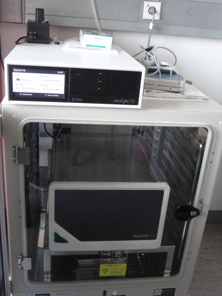
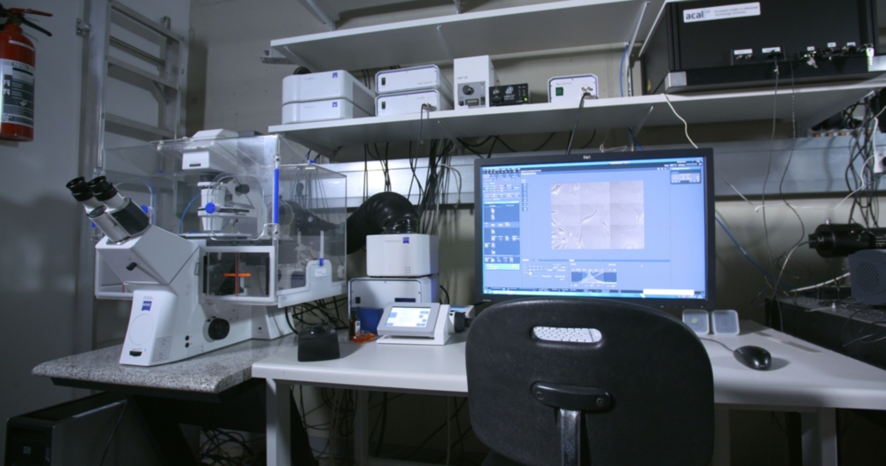
Fully automated epifluorescence microscope, located inside an incubator for longterm observation of living cells in up to six vessels simultaneously.
Real-time monitoring and analysis of multiple integrated live-cell assays.
Equipment: 4x / 10x / 20x phase contrast objectives, green and red fluorescence filters.
Applications (selection):
Apoptosis, Cytotoxicity, Proliferation, Spheroids, Scratch Wound Migration and invasion, chemotaxis, Dilution cloning, transfection efficiency, Cell-by-Cell Analysis
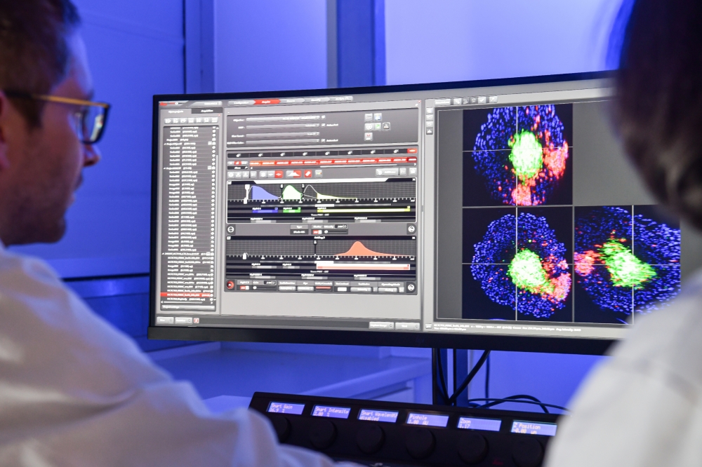
Next generation confocal laser scanning microscope with super resolution and high speed modus. The 32 PMT Array detector of the Airyscan 2 increases the lateral resolution to 120 nm (lateral) and 350 nm (axial). The refined multiplex modus allows to optimize acquisition speed (up to 47 fps), bleaching/phototoxicity and resolution according to individual experimental demands in an intelligent manner.
The inverted microscope is equipped with 6 excitation lasers (405 nm, 445 nm, 488 nm, 514 nm 561 nm, 640 nm), incubation chamber, CO2 unit, Definite Focus 3 and AI Sample finder.
Additional software modules: FCS, FRET, Physiology, 3d-rendering
Objectives:
Plan-Apochromat 63x / 1.4 Oil DIC
LD LCI Plan-Apochromat 40x / 1.2 Multi-Immersion
C-Apochromat 40x / 1.2 W
Plan-Apochromat 20x / 0.8
EC Plan-Neofluar 10x / 0.3
Applications:
Confocal imaging with super resolution. Gentle live cell imaging with high speed and sensitivity. FCS, FRET, FRAP, photoactivation, Colocalization
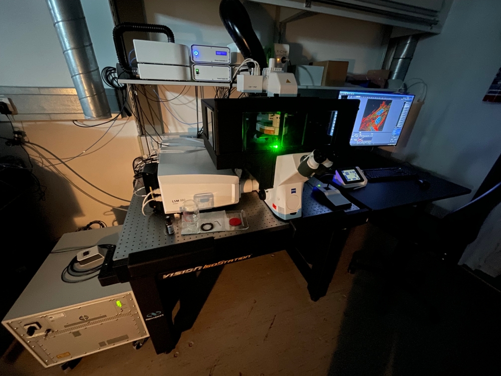
Inverted confocal laser scanning microscope equipped with 4 lasers providing excitation wavelengths of 405 nm, 458 nm, 488 nm, 514 nm, 561 nm and 633 nm.
Three detectors including a spectral detector for Lambda-mode, T-PMT for DIC images and a large incubation unit.
Objectives:
Plan-Apochromat 63x / 1.4 Oil DIC
EC Plan-Neofluar 40x / 1.3 DIC
Plan-Apochromat 20x / 0.8
EC Plan-Neoflura 10x / 0.3
Applications: 3d z-stacks in high resolution, live cell imaging, FRET, FRAP, Photoactivation, colocalization, linear unmixing
Inverted epifluorescence microscope equipped with ApoTome, Definite Focus 1, Colibri LED light source and a large incubation unit.
CCD camera: Axiocam MRm
Objectives:
Plan-Apochromat 63x / 1.4 Oil DIC
LD Plan-Neofluar 40x / 0.6 Korr Ph2
Plan-Apochromat 20x / 0.8
EC Plan-Neoflura 10x / 0.3
Applications: Long-term live cell imaging, multipositioning, 3d z-stacks with Apotome
Inverted confocal Spinning-Disk Microscope equipped with Yokogawa CSU-X1 Spinning Disk Unit, Laser-TIRF3, Definite Focus 2, a large incubation unit and UGA-42-Firefly point scanning device (Rapp Optoelectronics) for photomanipulation experiments.
6 acquisition lasers: 405 nm, 445 nm, 488 nm, 514 nm, 561 nm, 638 nm
2 photomanipulation lasers: 473 nm, 515 nm
CCD cameras Axiocam 503 mono (2x)
EMCCD camera: Photometrix Evolve 512
Objectives:
Plan-Apochromat 100x / 1.46 Oil DIC
Plan-Apochromat 63x / 1.4 Oil DIC
Plan-Apochromat 40x / 1.4 Oil DIC
LD LCI Plan-Apochromat 25x / 0.8 Imm Korr DIC
Plan-Apochromat 20x / 0.8
Plan-Apochromat 10x / 0.45
Applications: Fast confocal long term live cell imaging, multipositioning, fast 3d z-stacks, TIRF microscopy, simultaneous acquisition of two fluorescent channels in dual camera mode, FRET, FRAP, photoactivation, Colocalization.
Inverted TIRF microscope equipped with three lasers (488 nm, 561 nm, 640 nm) and EMCCD camera (Andor iXon Ultra 897)
Objectives:
CFI Apochromat 10x / 0.25
CFI Apochromat TIRF 100x / 1.49 Oil
Applications: Fast confocal live cell imaging, fast 3d z-stacks, TIRF microscopy, FRET, FRAP, Photoactivation, Colocalization
Applications: TIRF Microscopy, single molecule tracking
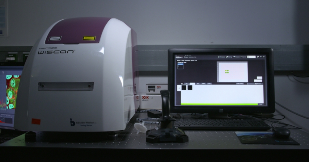
High content screening microscope for the automated acquisition of images in up to 1536-well plates in five channels (TL, blue, green, red, far red) equipped with live-cell package.
Objectives: 10x, 20x, 40x
Analysis software suite: WiSoft Minerva
Applications: High content screening, phenotypic screening, Live cell imaging
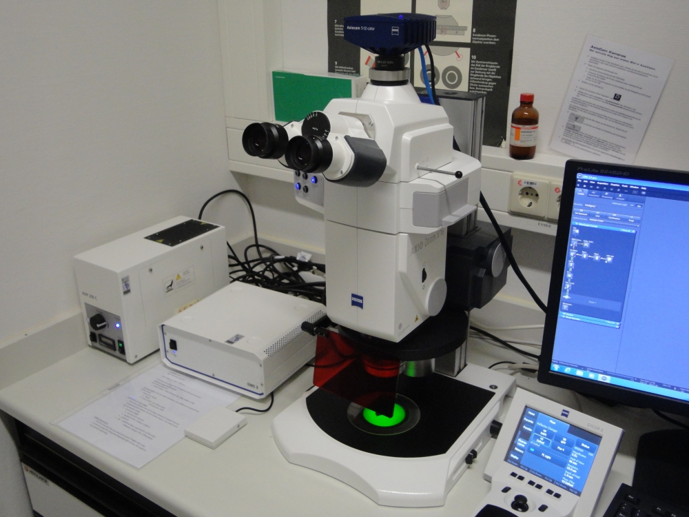
Fully automatized zoom microscope equipped with fluorescence in three channels (blue, green, red), 16x zoom optics and an adjustable field of view.
Objective: Plan Apo Z 0.5x / 0.125
Camera: AxioCam 512 Color
Applications: Imaging of large samples/fields of view, preparative microscopy
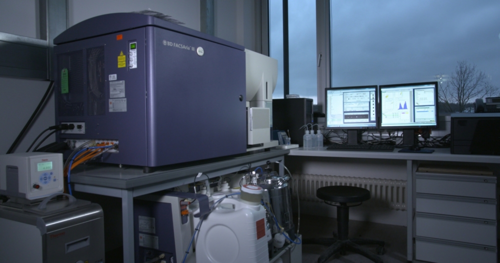
BD FACS Aria III, equipped with 4 Lasers (405 nm, 488 nm, 561 nm, 633 nm), 16 fluorescent detectors, FSC, SSC, a cooling unit and multiwell plate sorting option.
The system allows sorting of up to four populations simultaneously.
Inverted epifluorescence microscope equipped with Sutter DG-4 excitation light source and improvision orbit emission filter wheel for fast image acquisition.
The microscope has also a eppendorf FemtoJet microinjection station and a large incubation unit available.
Objectives: 10x, 20x, 25x, 40x, 63x,
Applications: Live cell imaging, FRET, microinjection

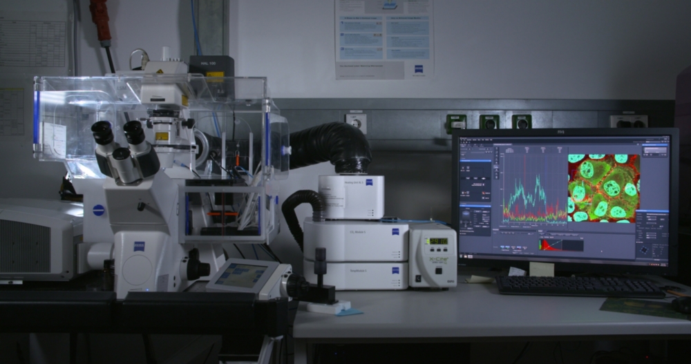
High-end confocal point scanning system for single- and multiphoton microscopy, inverted stand with large incubation unit. The Multiphoton unit allows flexible multicolor imaging as well as label-free contrast images like Second or Third Harmonic Generation (SHG/THG) in highly scattering tissue and scaffold samples up to 1 mm deep. Additionally, the system offers flurescence lifetime-based functionality in combination with pulsed lasers. Examples are spectral separation of overlapping fluorophores, removal of autofluorescence and semi-quantitative measurements using biosensors (protein-protein interaction, cell signaling, metabolic status etc.).
Multiphoton specifications:
Spectraphysics Insight X3 dual laser including 700-1300nm tunable line and 1045 nm fixed line.
4tune detection unit equipped with three spectrally tunable NDS HyD S detectors.
Single photon specifications:
Whitelight laser (pulsed, tunable wavelength 440nm - 790nm), 405 nm laser, AOBS, three HyD S detectors, detection range 410 – 850 nm
Objectives:
HCPL FLUOTAR 10x/0,30
HC PL APO 20x/0,75 CS2
HC FLUOTAR L 25x/0,95W VISIR 0,17
HC PL IRAPO 40x/1,1 W CORR
HC PL APO 63x/1,3 GLYC CORR CS2
Location of the Technology Platform Cellular Analytics
Downloads
Contact

Stephan Eisler
Dr.Scientific staff
[Image: SRCSB]

Melanie Noack
Technical staff
[Image: SRCSB]